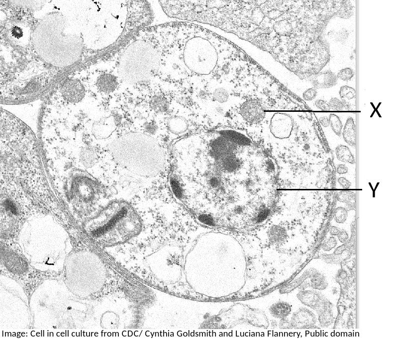
animal cell under electron microscope
Moreover because of their flexible nature they also facilitate movement. Animal cells Almost all animals and plants are made up of cells.
Animal Vs Plant Cells Similarities Differences Chart And Examples Rs Science
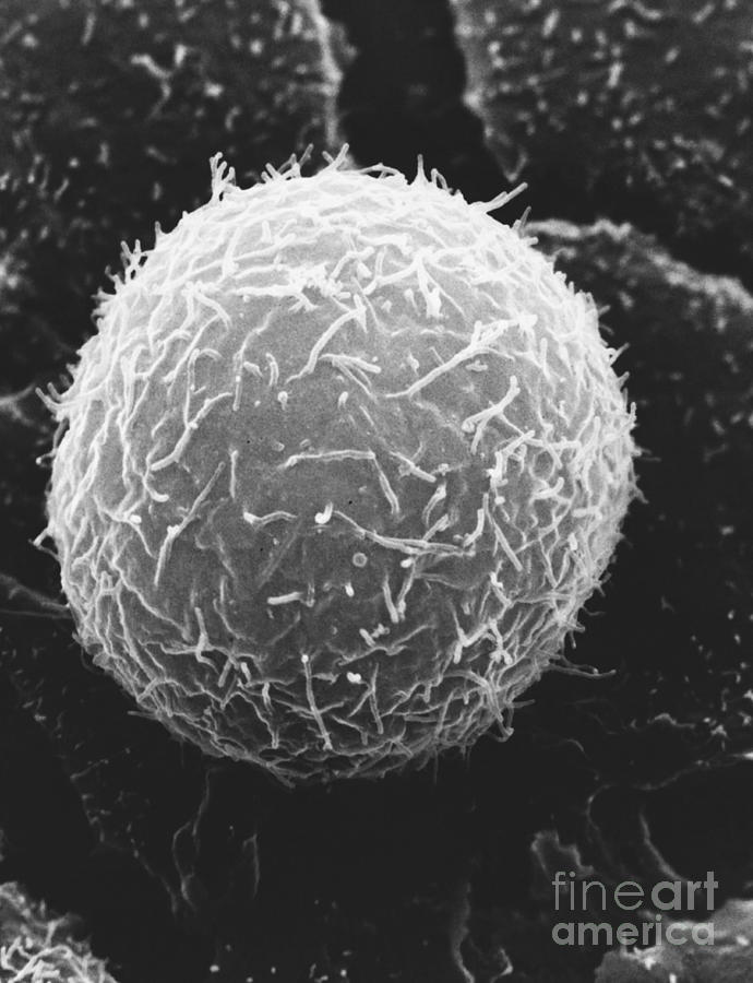
A capability for scanning electron microscopy of wet biological specimens is presented.

. With the unaided eye one can only see exceptionally large cells such as the human ovum which has a diameter of 100 µm. Most cells both animal and plant range in size between 1 and 100. Rabies seen here under a microscope is an often fatal viral disease that a generalised animal cell as observed under an electron microscope.
The TEM has revealed structures in cells that are not visible with the light microscope. Again microfilaments and intermediate. Viewing Animal Cells under a microscope.
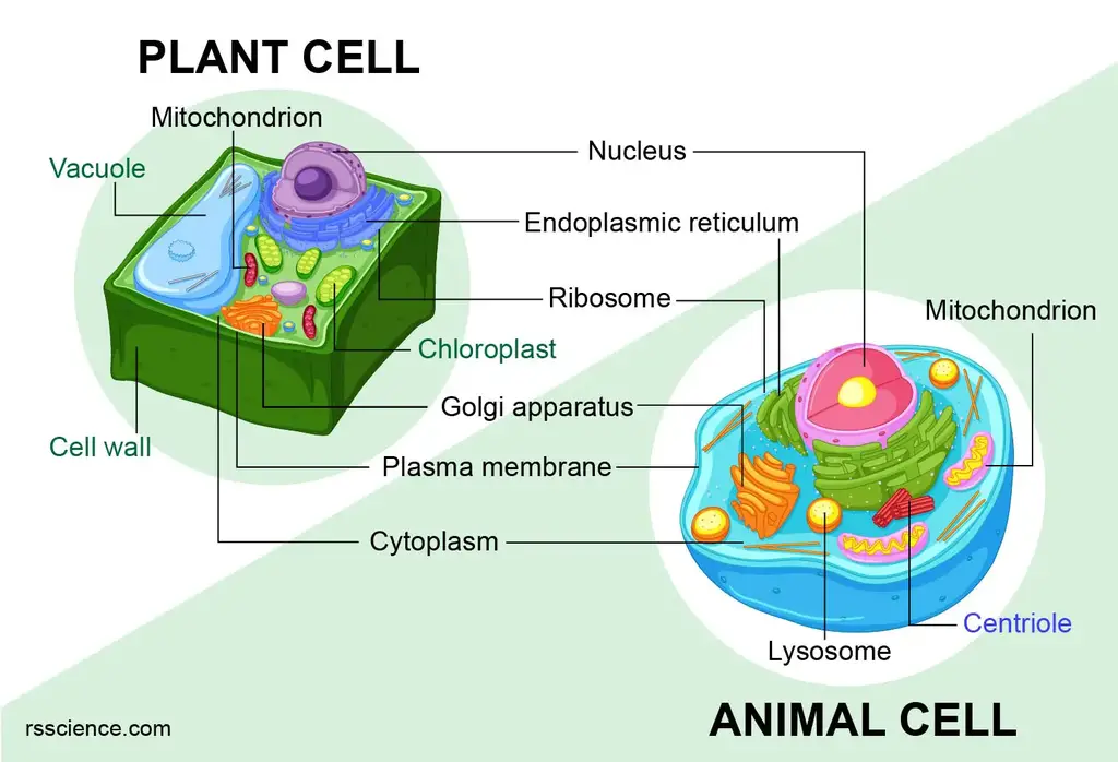
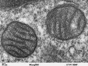
Cell Structures as Seen under the Light and Electron Microscope Cell Structure under Light Microscope. The animal cell is more fluid or elastic or malleable in structure. Eukaryotic cells contain organelles that are attached to membrane including endoplasmic reticulum ribosome mitochondria Golgi apparatus and more.
You will find the endoplasmic reticulum and mitochondria in the cytoplasm of the endothelium cell under the electron microscope. As for seeing electrons under any microscope in general i would say we have come as close to it as scientifically and technically possible with. Therefore we must use a microscope to visualize cells in a tissue.
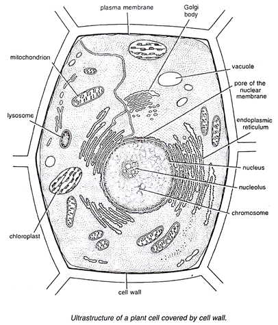
The cell membrane is what. Beneath a plant cells cell wall is a cell membrane. Heres a diagram of a plant.
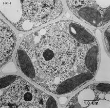
SEMs are often used at lower magnifications up to 30000. In order to view the organelles an electron microscope is needed. Below the basic structure is shown in the same animal cell on the left viewed with the light microscope and on the right with the transmission electron.
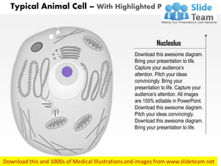
Almost all animals and plants are made up of cells. Animal cells have a basic structure. Animal cell under electron microscope.
Animal cells have a basic structure. These are also called Suicide bags or Death bags of the cell Fig. The structures within the cell are referred to as organelles.
The limit of resolution of a SEM is lower. An animal cell also contains a cell membrane to keep all the organelles and cytoplasm contained but it lacks a cell wall. Below the basic structure is shown in the same animal cell on the left viewed with.
The electron microscope Electron. A membrane that is transparent to electrons protects the fully. Animal cells range in size from.
Ultrastructure Of Cells 1 2

Ultrastructure

Cell Micrographs Bioninja
Topic 1 2 Ultra Structure Of Cells Amazing World Of Science With Mr Green

Magnification Questions Doc Cell Magnification Fig 1 2 1 Below Shows An Animal Cell 5m Fig 1 2 1 Diagram Showing The General Structure Of An Course Hero
Mitochondrion Wikipedia
What Is A Diagram Of A Plant And Animal Cell Under An Electron Microscope Quora

Eukaryotic Cells Under The Microscope 2 1 6 Ocr A Level Biology Revision Notes 2017 Save My Exams

Animal Cell Definition And Examples Biology Online Dictionary

Animal Cell Tem Stock Image C015 0851 Science Photo Library
Typical Animal Cell Sem Photograph By David M Phillips Fine Art America

1 2 Difference Between Plant And Animal Cells Cells As The Basic Units Of Life Siyavula

Animal Cell Tem Stock Image C025 2692 Science Photo Library

Animal Cell Plant Cell Diagram Cell Diagram Animal Cell Structure

Tem Of Animal Cell Stock Image G450 0055 Science Photo Library
Illustrate Only A Plant Cell As Seen Under Electron Microscope How Is It Different From Animal Cell Sarthaks Econnect Largest Online Education Community
The Cell Form 1 Biology Notes Easy Elimu
A Typical Animal Cell As Seen In An Electron Microscope Medical Ima

Microscopic Animal Cells Images Kuhn Photo